ChinSoo, W., Georges, D.
Thoracic Medical Department, Eric Williams Medical Sciences Complex, North Central Regional Health Authority, Trinidad and Tobago
Corresponding author
Dr. D. Georges
Thoracic Medical Unit
Eric Williams Medical Sciences Complex
North Central Regional Health Authority
Champs Fleurs
Trinidad and Tobago
E-mail: [email protected]
Copyright: This is an open-access article under the terms of the Creative Commons Attribution License which permits use, distribution, and reproduction in any medium, provided the original work is properly cited.
Introduction
Pulmonary hypertension (PH) is a condition that is being currently diagnosed much more frequently in comparison to the past. This pathophysiological condition is defined as a mean pulmonary arterial pressure (PAP) greater than 20 mmHg measured ideally by right heart catheterization (RHC). In the past, the threshold for PAP used to be 25 mmHg; however, since increased mortality has been found even in patients who have mild PH (PAP >19 mm Hg), the guidelines have been updated internationally. Such patients require detailed workup inclusive of mandatory RHC. Echocardiography, specifically transthoracic echocardiography (TTE), can give a provisional diagnosis. In our experience, there have been challenges in obtaining these procedures in the local pulmonary medicine setting. This has resulted in the diagnosis of PH being performed with a computed tomography pulmonary angiogram (CTPA), a trend which is inappropriate and not in keeping with evidence-based medicine.
Case Reports
We describe two case scenarios encountered in our settings in order to highlight the management of PH in a resource-constrained setting.
Case 1 was an 80-year-old female who was referred for investigation of pulmonary hypertension following chronic obstructive pulmonary disease (COPD). She presented with palpitations and was an ex-smoker of 40 pack-years. She had a computerised tomography of pulmonary artery (CTPA) scan which revealed a dilated pulmonary trunk measuring 35 mm diameter, suggestive of PH.
Case 2 was a 68-year-old female who was referred for investigation of PH following open-heart surgery. Chest X-ray revealed cardiomegaly with a prominent pulmonary artery and small left-sided pleural effusion. She was Day-19 post mitral valve replacement.
Both cases are representative of typical referrals received in our setting for investigation and management of pulmonary hypertension PH. The referral letters were brief, did not contain any detailed work-up and the clinical information was inadequate. Although referred by cardiologists to pulmonologists, there was no mention of right ventricular systolic pressure (RVSP) measured by echocardiogram. Hence, both referrals were returned to the referring physicians with requests for more clinical information, including RVSP.
In Case 1, a transthoracic echocardiogram was done which revealed normal structure and function of the tricuspid and pulmonary valves, mild tricuspid regurgitation (TR), an RVSP of 15 mmHg (normal) and mild concentric left ventricular hypertrophy (LVH) with a preserved ejection fraction (EF) of 60-65%.
With respect to Case 2, the referring physicians did not follow-up the patient with the pulmonologists. Pulmonary hypertension is most commonly caused by left heart disease and this patient had mitral valve disease. The patient also had a mitral valve replacement and was treated for the underlying condition. It would have been interesting to know both the initial RSVP and that after the mitral valve replacement and whether a right heart catherization RHC had been done prior to surgery. However, the information on this patient remained inadequate.
Discussion
Diagnosis of Pulmonary Hypertension
The transthoracic echocardiogram (TTE) is an important non-invasive screening tool to diagnose PH. This modality uses many different parameters derived from well-established data (normal as well as abnormal) to predict the likelihood of Pulmonary hypertension.1
In the local setting, a provisional diagnosis of PH is made when the right ventricular systolic pressure (RVSP) is more than 40 mmHg on echocardiogram. However, Strange et al. have opined that this threshold underestimates the prognostic impact since an RVSP of 30 mmHg has been associated with an increased risk of mortality.2 Hence, right heart catheterization (RHC) may be mandatory for establishing the diagnosis of PH, particularly when therapeutic strategies are being considered.3
In the past, a definitive diagnosis of pulmonary hypertension PH was made when the mean PAP was greater than 25 mmHg at rest on RHC. Later, it was found that the mortality risk increased even with a PAP between 20 mmHg and 25 mmHg. Therefore, the diagnosis of PH is now defined as a PAP of 20 mmHg and more, which is 2 standard deviations above the normal mean value of 14.0 ± 3.3 mmHg.4,5
The 6th World Symposium on Pulmonary Hypertension defined three haemodynamic profiles of PH:
- Isolated pre-capillary PH
- Combined pre-capillary and post-capillary PH
- Isolated post-capillary PH
The haemodynamic profiles of the categories of PH are summarized in Table 1. (6)
Table 1. Haemodynamic profiles of pulmonary hypertension (Source Ref 6)
| Classification | Mean pulmonary artery pressure (PAP) | Pulmonary capillary wedge pressure
(PCWP) |
Pulmonary vascular resistance
(PVR) |
| Isolated pre-capillary PH | >20 mmHg | <15 mmHg | >3 WU |
| Combined pre- and post-capillary PH | >15 mmHg | >3 WU | |
| Isolated post-capillary PH | >15 mmHg | <3 WU |
WU, Wood Units.
Treatment of PH
Over the last 50 years, there has been an increase in the treatment options available for PH. Phosphodiesterase 5 inhibitors (PDE-5i), such as sildenafil and tadalafil, block the degradation of cyclic GMP. Synthetic prostacyclins, such as epoprostenol, are potent vasodilators with short half-lives that are usually given parenterally in the inpatient setting. The endothelin receptor antagonists (ERAs), such as bosentan and abrisentan, are newer agents and decrease the serum endothelin concentrations. Other newer agents are rociguat (which enhances cGMP production) and selexipag (which is a prostacyclin agonist).
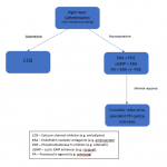
In terms of specific treatment, monotherapy is not advised, even in mild PH, except for a few well-defined clinical scenarios. In the event that a patient improves clinically, stepping down of therapy is not routine. Combination therapy using drugs with different mechanisms of action has been proven to be more effective than single drugs, at least in the initial stages. For difficult cases, transition between therapies should only be done with careful monitoring in an expert centre.6 The treatment algorithm in ideal conditions is summarized in Figure 2.
As more patients use these new treatment combinations, we will be able to gather more data on long-term effects on disease progression, survival and health related quality of life.6,8
Issues in diagnosing and managing PH
Is it a best practice to diagnose PH exclusively based on CTPA and start sildenafil?
Jaramillo et al. advocated a CT approach to the diagnosis of pulmonary hypertension, although this was a recommendation from the radiologists. It involves identifying an enlarged pulmonary artery diameter (>29 mm) which is usually larger than that of the ascending aorta at the same level. This diameter must be measured in the axial plane at the bifurcation, orthogonal to the long axis of the pulmonary artery.8
In our first case, the CTPA showed a dilated pulmonary truck measuring 35 mm. However, TTE done on the same patient showed a RVSP at 15 mmHg which is normal. This demonstrates the lack of correlation between the two investigations. While echocardiograms can over-estimate or under-estimate the RVSP, it may be only used provisionally. On the other hand, there are no established guidelines which recommend the use of CTPA as a diagnostic for pulmonary hypertension. Therefore, using a CTPA alone to diagnose pulmonary hypertension and starting medications on that basis may not be considered best practice.
In our setting, it is easier to obtain a CTPA rather than an echocardiogram or a right heart catheterisation RHC. Even though an echocardiogram is available in the public service, patients routinely wait months to get an appointment and even longer to get the report. Also, most patients cannot afford to have an echocardiogram done privately.
With respect to treatment, the current guidelines suggest the use of combination therapy even at disease onset, however, synthetic prostacyclins and endothelin receptor antagonists are not affordable for the majority of patients seen in the public healthcare system.
The most common cause of pulmonary hypertension is left heart disease, for which the appropriate treatment is to treat the underlying disease. If PH-specific therapy is considered, the cause of the pulmonary venous hypertension should first be optimally treated, the pulmonary capillary wedge pressure (PCWP) should be normal or minimally elevated and the transpulmonary gradient (TPG) and pulmonary vascular resistance (PVR) should be significantly elevated. Such treatment should be undertaken with great caution as it may increase fluid retention, left-sided filling pressures, pulmonary oedema and may result in clinical deterioration.
Similarly, in pulmonary hypertension related to chronic lung disease and hypoxaemia, the underlying condition should first be optimally treated and the TPG and PVR should be significantly elevated. Otherwise, PH-specific therapy may result in worsening hypoxaemia and clinical deterioration.
Conclusion
There are many issues in diagnosing and managing pulmonary hypertension, some are unique to low-resource settings. However, even in low resource settings, diagnosing and treating PH exclusively based on CTPA findings is not evidence-based and should be discouraged. Right heart catheterizations RHCs and TTEs, though available, are difficult to access by the majority of patients in this setting. However, public healthcare settings must increase the capacity of transthoracic echocardiogram TTE, by increasing human and material resources in order to optimally diagnose and manage PH patients.
With respect to management, current guidelines suggest the use of combination therapy even at the onset of the disease. However, synthetic prostacyclins and endothelin receptor antagonists are not yet on the National Formulary and are very expensive. Thus, they are unaffordable for the majority of patients seen in public healthcare settings and consideration must be given to provide them in public hospitals.
It is vital that the treatment approach to pulmonary hypertension includes a multidisciplinary team targeting both the medical and social aspects of this chronic, debilitating disease.
Ethical Approval:
This was a commentary on the state of the management of pulmonary hypertension in Trinidad and Tobago. There was no direct patient contact and the information was obtained from the referral letters.
Informed Consent: Not applicable
Funding statement: No funding was needed as no research was carried out. Reference articles for literature review were widely accessible.
Authors’ contributions: Dr Chin Soo wrote the skeleton draft. Both authors did research to elaborate and expand on the original version. Formatting according to CMJ guidelines was done by both authors.
Conflict of interest: None to declare. Neither author is associated with any pharmaceutical companies.
References
- Frost A, Badesch D, Gibbs J, Gopalan D, Khanna D, Manes A et al. Diagnosis of pulmonary hypertension. European Respiratory Journal. 2019;53(1):1801904.
- Strange G, Stewart S, Celermajer D, Prior D, Scalia G, Marwick T et al. Threshold of pulmonary hypertension associated with increased mortality. Journal of the American College of Cardiology. 2019;73(21):2660-2672.
- “2015 ESC/ERS Guidelines for the diagnosis and treatment of pulmonary hypertension. The Joint Task Force for the Diagnosis and Treatment of Pulmonary Hypertension of the European Society of Cardiology (ESC) and the European Respiratory Society (ERS).” Galiè N, Humbert M, Vachiery J-L, et al. Eur Respir J 2015; 46: 903–975. European Respiratory Journal. 2015; 46 (6): 1855-1856.
- Kolte D, Lakshmanan S, Jankowich M, Brittain E, Maron B, Choudhary G. Mild pulmonary hypertension is associated with increased mortality: A systematic review and meta‐analysis. Journal of the American Heart Association. 2018;7(18).
- Rajpal S. The 6th World Symposium on PH: Hemodynamic Definitions and Updated Clinical Classification of PH (Part 1) – American College of Cardiology [Internet]. 2020 [cited 30 November 2019]. Available from: https://www.acc.org/latest-in-cardiology/articles/2019/10/30/08/08/the-6th-world-symposium-on-ph-part-1
- Condon D, Nickel N, Anderson R, Mirza S, de Jesus Perez V. The 6th World Symposium on Pulmonary Hypertension: what’s old is new. 2019; 8: 888.
- Davies R, Howard L. Pulmonary vascular disease: pulmonary thromboembolism and pulmonary hypertension. Medicine. 2016;44(4):255-262.
- Aluja Jaramillo F, Gutierrez F, Díaz Telli F, Yevenes Aravena S, Javidan-Nejad C, Bhalla S. Approach to Pulmonary Hypertension: From CT to Clinical Diagnosis | RadioGraphics [Internet]. Pubs.rsna.org. 2018. Available from: https://pubs.rsna.org/doi/10.1148/rg.2018170046.


